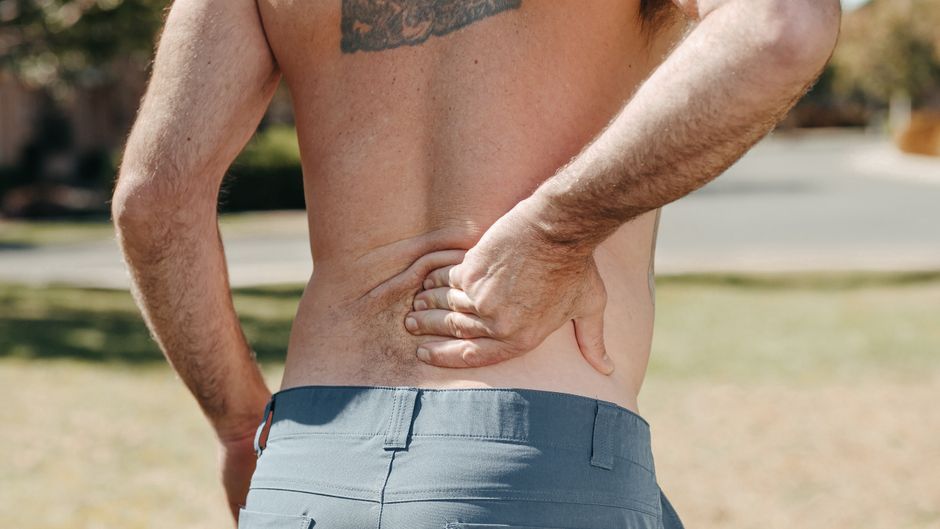
A compressed nerve in the lower back often causes more than a dull, local ache. Pain that shoots or burns from the low back or buttock into one leg, along with tingling, numbness, or weakness, is more suggestive of nerve irritation than a simple muscle strain.
Quick ways to tell the difference:
A herniated disc, arthritis-related bone spurs, or narrowing around the spinal nerves can cause this pressure. The medical term is often lumbar radiculopathy; "sciatica" is the common term when symptoms travel through the buttock and down the leg.
I am Sonny Dosanjh, M.D., a board-certified Physical Medicine and Rehabilitation physician and fellowship-trained pain specialist who evaluates compressed nerve in lower back symptoms, sciatica, sports injuries, and chronic spine pain. At Medici Orthopaedics & Spine, my focus is identifying the true pain source and building the least invasive, least drug-dependent path back to movement and daily life.

The lower spine consists of five lumbar vertebrae separated by shock-absorbing intervertebral discs. Thirty-one pairs of spinal nerves branch off the spinal cord and exit through small bony tunnels called neural foramina. When surrounding structures intrude into these narrow pathways, mechanical compression and chemical irritation compromise normal nerve function.

When an intervertebral disc tears or herniates, its gel-like inner core (nucleus pulposus) leaks into the spinal canal or lateral recess. This displacement directly presses against the traversing or exiting nerve root while releasing inflammatory cytokines. Our detailed lumbar radiculopathy definition guide explains how this combination of mechanical pinching and biochemical inflammation triggers true radiculopathy.
According to a clinical review of lumbosacral radiculopathy, the lifetime prevalence of low back pain ranges from 60% to 90% in the general population, with 5% to 10% of those individuals developing radiculopathy. Recognizing whether your discomfort is a muscular spasm or true neural impingement is essential for preventing long-term nerve irritation.
Sciatica is a descriptive term rather than an independent medical diagnosis. It refers to symptoms that travel along the expansive sciatic nerve, formed by the convergence of the L4, L5, S1, S2, and S3 nerve roots. When any of these individual roots become pinched in the lower back, symptoms project down the buttock, the back of the thigh, and into the foot. Learn more about how these nerve pathways interact in our guide to sciatica and lumbar radiculopathy.
In older adults, neural foraminal stenosis often stems from degenerative osteophytes (bone spurs) and facet joint enlargement rather than acute disc tears. As outlined in the lumbar spinal stenosis characteristics, this progressive narrowing affects roughly 11% of older individuals in the United States, rising to 10.8% in adults aged 70 to 79.
Low back pain is the leading cause of disability worldwide and the second most common reason for missed work. Differentiating localized muscular pain from nerve root compression ensures you receive appropriate care early on.
| Characteristic | Mechanical Muscle Strain | Compressed Spinal Nerve (Radiculopathy) |
|---|---|---|
| Pain Quality | Dull, achy, sore, throbbing | Sharp, electric, shooting, burning |
| Location | Localized to the lumbar region/flanks | Radiates below the knee into the calf, ankle, or foot |
| Sensory Changes | None (sensation remains fully intact) | Numbness, tingling, "pins and needles" (paresthesia) |
| Motor Impact | Stiffness, guarding, limited spinal range | Weakness in specific muscles, foot drop, diminished reflexes |
| Cough/Sneeze Effect | Minimal change | Sharp, sudden spikes in shooting pain (Valsalva aggravation) |
| Positional Triggers | Aggravated by bending or lifting | Aggravated by prolonged sitting or spinal extension |
If your discomfort is confined to the lumbar muscles without radiating sensations, exploring treatment for underlying lower back pain can help restore spinal mechanics and resolve soft tissue tension.
A trapped lumbar nerve produces distinctive neurological sensations:
For further insight into what generates these severe spikes, read our resource on identifying sharp nerve pain in your lower back.
Prolonged or severe nerve compression interrupts motor signaling to the lower extremities:
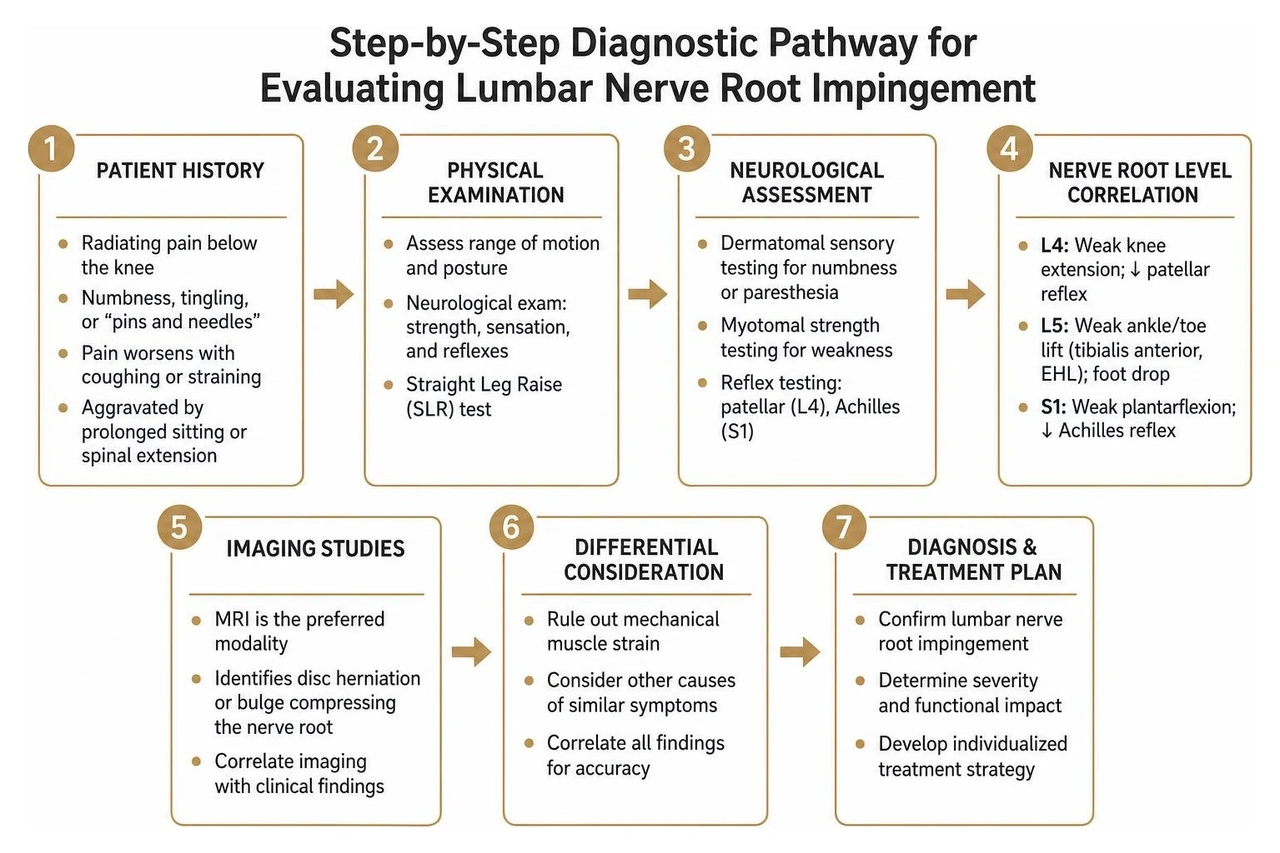
A variety of spinal conditions can cause neural impingement, as described in Cleveland Clinic's review of pinched nerve mechanisms:
During an in-office physical examination, we use provocative orthopedic tests to assess nerve root tension:
If red flags appear or symptoms persist beyond 4 to 6 weeks, non-contrast lumbar MRI serves as the diagnostic standard to visualize soft tissue structures, disc herniations, and neural exit pathways. Electromyography (EMG) and nerve conduction studies (NCS) can also measure electrical signal velocity to localize specific root entrapment.
While most lower back nerve compression cases are safely managed with conservative care, Cauda Equina Syndrome is a true orthopedic emergency requiring decompression within 24 to 48 hours to prevent permanent dysfunction:
Fortunately, roughly 50% of acute lower back pain episodes improve within 1 to 2 weeks, and up to 90% resolve within 6 to 12 weeks with conservative management. At Medici Orthopaedics & Spine, we emphasize a tiered care pathway designed to relieve nerve compression without unnecessary surgery. Discover practical options in our breakdown of the best treatment for nerve pain in the lower back.
First-line conservative therapies include:
When conservative modalities need additional support, image-guided interventional procedures deliver targeted relief directly to irritated neural tissue:
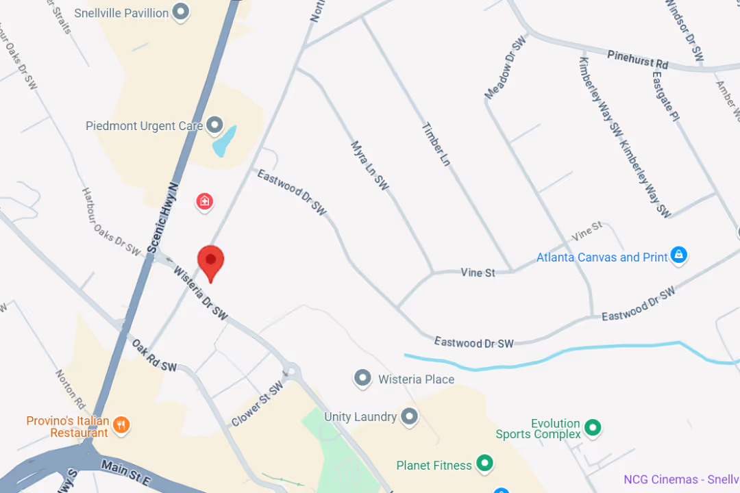
If you are located in Metro Atlanta, explore specialized nerve pain in lower back treatment in Snellville and our surrounding clinics.
Surgery is generally considered only when conservative and interventional options have been exhausted over 6 to 12 weeks, or when progressive motor deficits develop.
Review our guidance on when to see an orthopedic spine specialist for lower back pain to determine the best timing for a specialist consultation.
Both conditions cause leg discomfort during walking, but their underlying mechanisms differ:
Most mild-to-moderate compressed nerves improve within 4 to 8 weeks through conservative management, targeted physical therapy, and activity adjustments. As localized swelling subsides, the nerve root recovers normal conduction. However, if compression is accompanied by persistent motor deficits, early interventional care helps prevent chronic neuropathic changes.
Living with compressed nerve symptoms in your lower back can disrupt your daily routine, sleep, and overall mobility. Fortunately, identifying the precise mechanical and neurological source of your symptoms allows for targeted, non-surgical relief.
At Medici Orthopaedics & Spine, Dr. Sonny Dosanjh and our multidisciplinary team combine advanced diagnostic evaluations, image-guided interventional pain management, regenerative therapies, and personalized rehabilitation. Our goal is to resolve nerve compression and restore your quality of life through the least invasive approaches available.
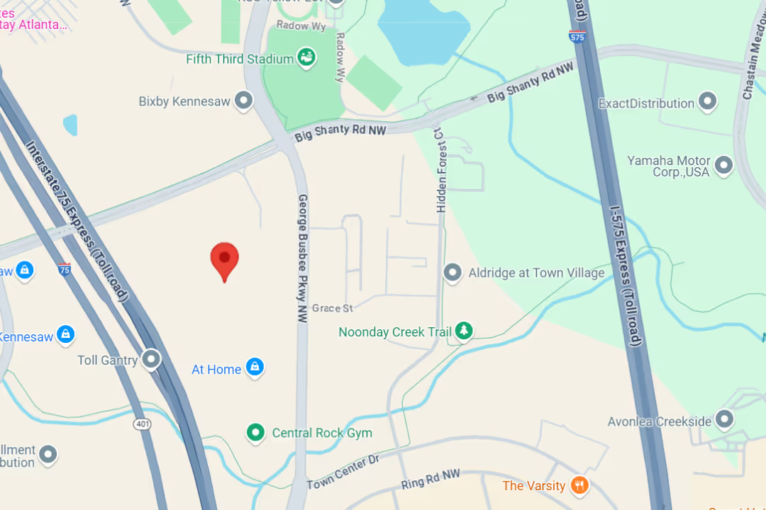
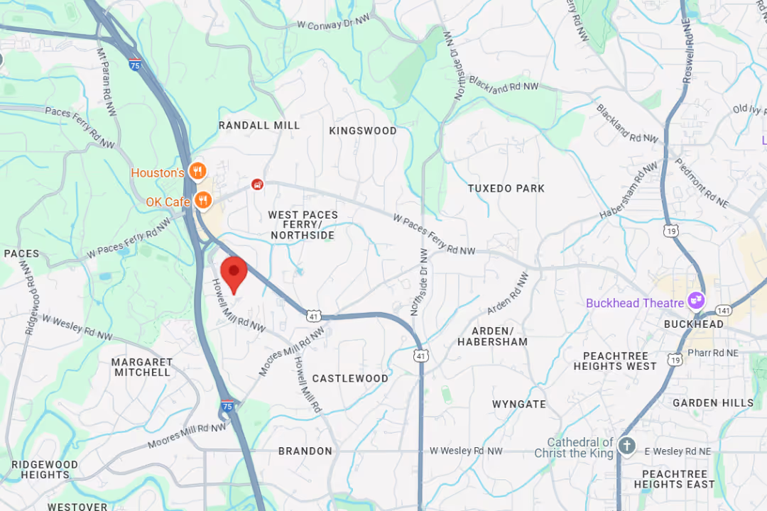
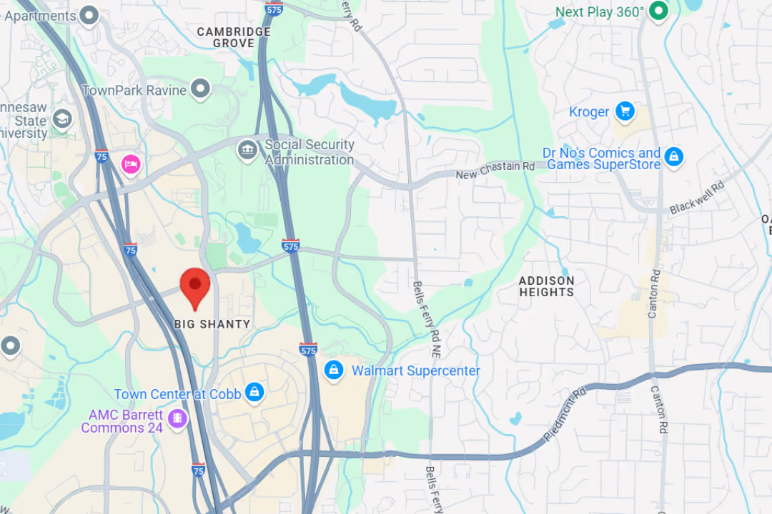
To begin your recovery, schedule a consultation for comprehensive lumbar radiculopathy care at any of our convenient locations across Metro Atlanta, including Snellville, Marietta, Kennesaw, and Buckhead.
At Medici, you’re more than your MRI.
We take time to hear your story, understand your pain, and create a plan that actually works for you.

Our team delivers specialist care at convenient locations across Metro Atlanta:
Get expert tips on injury recovery, pain relief, joint health, and movement strategies—straight from our Fellowship-Trained team.