The definition of lumbar radiculopathy is straightforward: it is a condition in which one or more nerve roots in the lower back become compressed or irritated, causing pain, numbness, tingling, or weakness that radiates down into the leg.
Quick answer:
Lumbar radiculopathy = compression or irritation of a nerve root in the lumbar (lower) spine, producing symptoms — pain, numbness, tingling, or muscle weakness — that travel along the path of that nerve into the buttock, leg, or foot.
Here is what sets it apart from simple back pain:
This condition is more specific — and more serious — than everyday back pain. While up to 90% of adults will experience low back pain at some point in their lives, only 5%–10% of those cases involve true radiculopathy. Yet when it does occur, it can be disabling enough to keep millions of people away from work and the activities they love.
I'm Dr. Sonny Dosanjh, M.D., a board-certified specialist in Physical Medicine & Rehabilitation and fellowship-trained in Multidisciplinary Pain Management at Emory University, with extensive clinical experience evaluating and treating the full spectrum of conditions covered by the definition of lumbar radiculopathy — from acute disc herniations to chronic nerve pain. In the sections below, I'll walk you through everything you need to know, from anatomy and diagnosis to the most effective, least invasive treatment options available in 2026.
To understand the clinical definition of lumbar radiculopathy, we must look beyond basic back soreness. In professional medical terms, as outlined in the Merck Manual Professional Edition, lumbosacral radiculopathy is a distinct neurological syndrome. It occurs when a disease process directly impairs the function of one or more nerve roots in the lumbosacral region of the spine.
This impairment is characterized not just by pain, but by objective neurological deficits. These deficits typically fall into two main categories:
In our clinics across Metro Atlanta, we often see patients who mistake generalized muscle spasms for nerve compression. True radiculopathy involves a structural or chemical threat to the nerve root itself, demanding a targeted diagnostic and therapeutic approach.
To visualize how this condition develops, it helps to understand the anatomy of the lower spine. The lumbar spine consists of five large vertebrae (labeled L1 through L5) stacked on top of one another, sitting just above the sacrum (S1).
Between these vertebrae lie intervertebral discs, which act as shock absorbers. At each spinal level, nerve roots branch off the spinal cord and exit the spinal column through small lateral openings called the neural foramina.
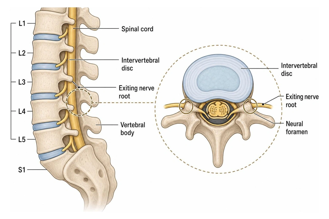
When we discuss the definition of lumbar radiculopathy in our Lumbar Radiculopathy (Pinched Nerve) Condition Guide, we are focusing on these exiting nerve roots. If a disc herniates (bulges outward) or if the neural foramina narrow due to bone spurs, the space available for the nerve root shrinks. The nerve becomes physically crowded, pinched, or stretched, triggering the radiating symptoms that travel down the leg.
It is a common misconception that nerve pain is solely caused by physical pinching. In reality, the pathophysiology of lumbar radiculopathy is a combination of mechanical compression and a complex chemical inflammatory cascade.
When an intervertebral disc tears or herniates, the soft inner core of the disc (the nucleus pulposus) leaks out. This material contains highly irritating inflammatory proteins. When these chemicals wash over the neighboring nerve root, they cause severe chemical irritation and swelling, even if the physical pressure on the nerve is minimal.
According to research on lumbar radicular pain in PMC, nerve roots are uniquely vulnerable to this double-whammy of mechanical and chemical irritation. Unlike peripheral nerves in your arms or legs, spinal nerve roots:
Because of this vulnerability, a stretch of just 10% to 15% of a nerve root's resting length is enough to cause severe neurophysiological dysfunction. This explains why even a minor disc bulge can sometimes cause agonizing, electric leg pain.
Because lower extremity pain can stem from several different sources, we must carefully differentiate true lumbar radiculopathy from other mimicking conditions.
| Feature | Lumbar Radiculopathy | Sciatica | Myelopathy | Peripheral Neuropathy |
|---|---|---|---|---|
| Primary Location of Pathology | Spinal nerve root (L1–S1) | Lower nerve roots (L4–S1) | Spinal cord (Cervical/Thoracic) | Distal peripheral nerves |
| Symptom Distribution | Single dermatome (one leg) | Back of the leg to the foot | Bilateral, diffuse, coordination loss | Symmetrical "glove-and-stocking" (feet/hands) |
| Reflex Changes | Hypoactive (reduced) in one reflex | Hypoactive (Achilles/Patellar) | Hyperactive (exaggerated), Babinski sign | Diminished bilaterally in feet |
| Common Cause | Herniated disc, stenosis | L4-S1 disc herniation | Spinal canal stenosis | Diabetes, metabolic issues |
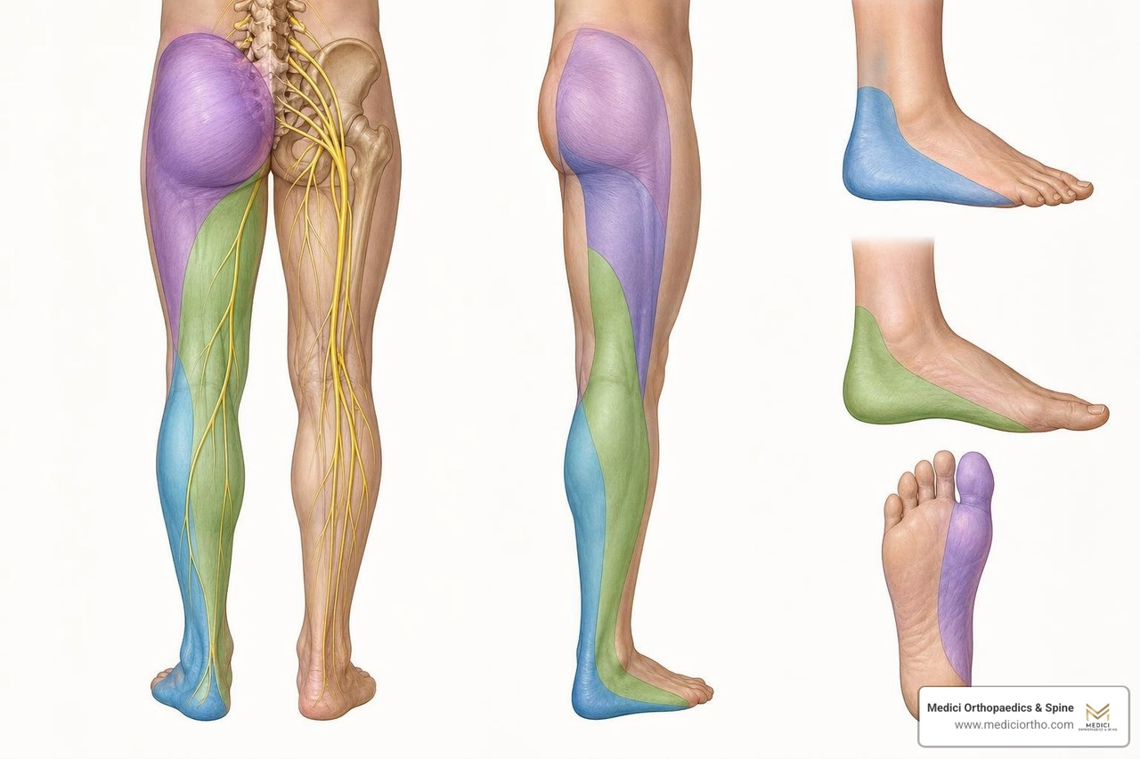
Understanding the specific pathway of your pain is one of the most powerful diagnostic tools we have. Because each spinal nerve root is responsible for sending signals to a specific patch of skin (a dermatome) and controlling specific muscles (a myotome), we can often pinpoint exactly where the nerve is pinched just by listening to your symptoms.
Before mapping the symptoms, let's look at what causes the compression in the first place, as detailed in our article on what's causing that sharp nerve pain in your lower back:
According to clinical guidelines on acute lumbosacral radiculopathy from UpToDate, the vast majority of cases affect the L4, L5, or S1 nerve roots. Here is how they present:
At Medici Orthopaedics & Spine, we believe that an accurate diagnosis is the foundation of successful recovery. We combine hands-on physical exams, advanced imaging, and targeted electrodiagnostic testing to build a precise map of your spinal health.
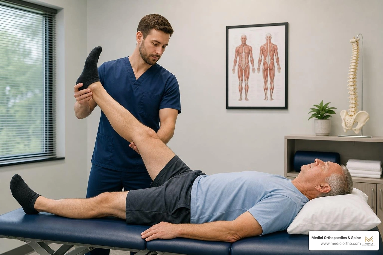
Our evaluation begins with specialized physical maneuvers designed to put gentle tension on the spinal nerves:
While a physical exam tells us how the nerve is behaving, advanced diagnostics show us why:
During your evaluation, we screen immediately for "red-flag" symptoms that indicate a medical emergency:
These symptoms point to Cauda Equina Syndrome, a rare but critical emergency where the bundle of nerves at the base of the spinal cord is severely compressed. If you experience any of these, seek emergency medical evaluation immediately.
If you are diagnosed with a pinched nerve, take comfort in this fact: the vast majority of patients do not need surgery. At Medici Orthopaedics & Spine, our philosophy is to utilize the most effective, least invasive treatments first.
For the first 4 to 6 weeks, we focus on reducing inflammation and supporting your body's natural healing processes:
If conservative care does not provide sufficient relief, we offer advanced, targeted treatments to bridge the gap before surgery is ever considered, as discussed in our FAQ on the best treatment for nerve pain in the lower back:
According to the landmark SPORT trial analyzed in the StatPearls Clinical Review of Lumbosacral Radiculopathy, both surgical and non-surgical patients show excellent, comparable recovery rates over the long term. However, surgery is indicated if you experience progressive muscle weakness, intractable pain that fails to respond to 6 weeks of conservative care, or red-flag emergencies.
When surgery is necessary, we prioritize minimally invasive options like a microendoscopic discectomy (MED), which uses a tiny tube and camera to remove the herniated portion of the disc with minimal disruption to the surrounding muscles.
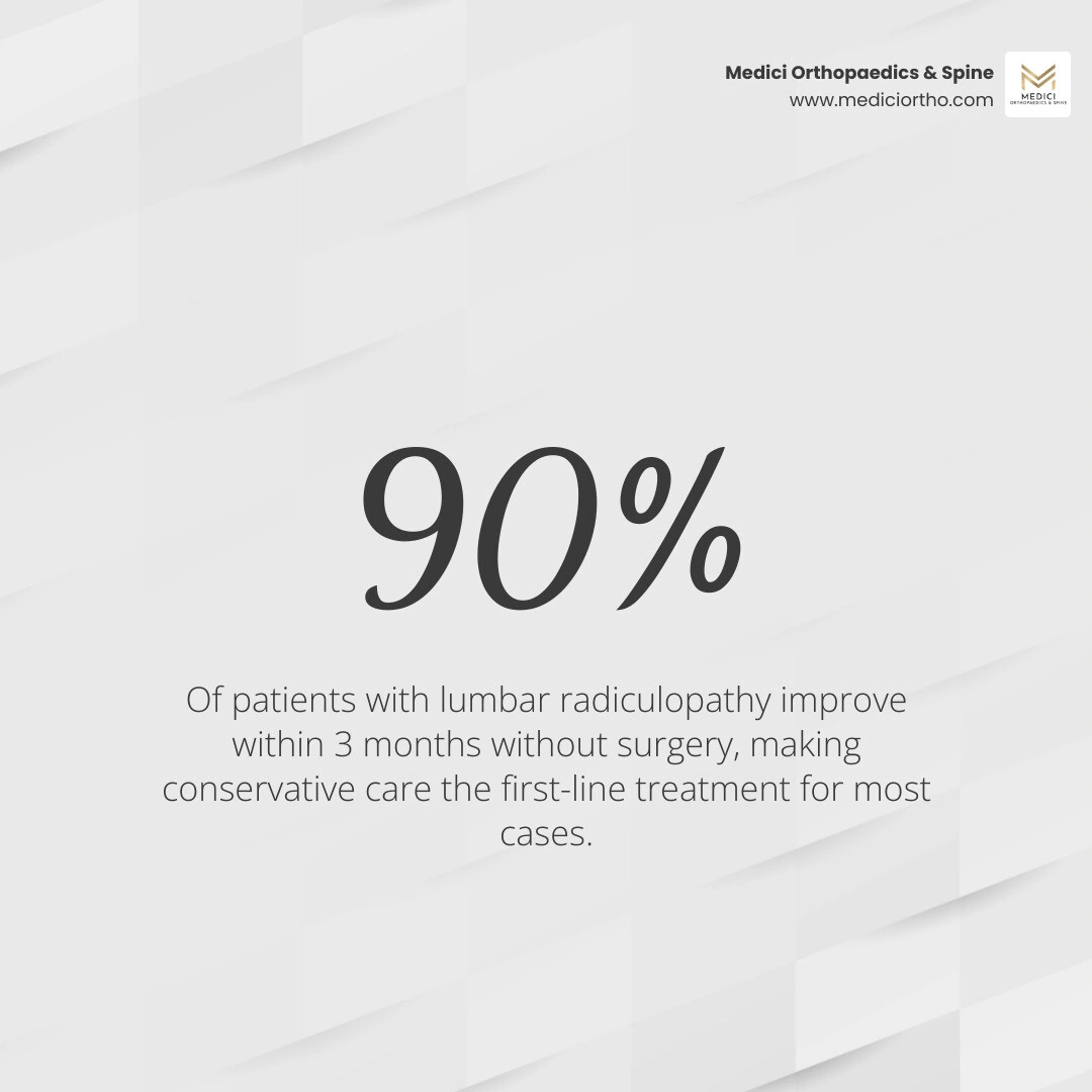
For most people, the prognosis is excellent. According to recovery data compiled by Healthline, roughly 50% of acute cases resolve within 1 to 2 weeks, and up to 90% of cases completely resolve within 6 to 12 weeks of starting conservative treatment. The body has an incredible ability to resorb herniated disc material over time, naturally relieving pressure on the nerve.
While you cannot stop age-related changes entirely, you can significantly reduce your risk of nerve compression by:
Surgery becomes an absolute, non-negotiable necessity only in the presence of "red-flag" symptoms, such as Cauda Equina Syndrome (loss of bowel/bladder control, saddle numbness) or sudden, progressive paralysis in your leg (like a rapidly worsening foot drop). For standard pain and mild numbness, a conservative trial of 4 to 8 weeks is always recommended first.
Understanding the definition of lumbar radiculopathy is the first step toward reclaiming your active lifestyle. While radiating leg pain can feel frightening and disruptive, it is a highly treatable condition that rarely requires invasive open surgery.
At Medici Orthopaedics & Spine, Dr. Sonny Dosanjh, M.D., and our dedicated medical team are committed to Optimally Restoring your Quality of Life with the most effective, least invasive, and least drug-dependent programs medically available today. We look at your body as a whole, combining physical therapy, advanced injections, hyperbaric oxygen therapy, and regenerative medicine to help you heal naturally and thoroughly.
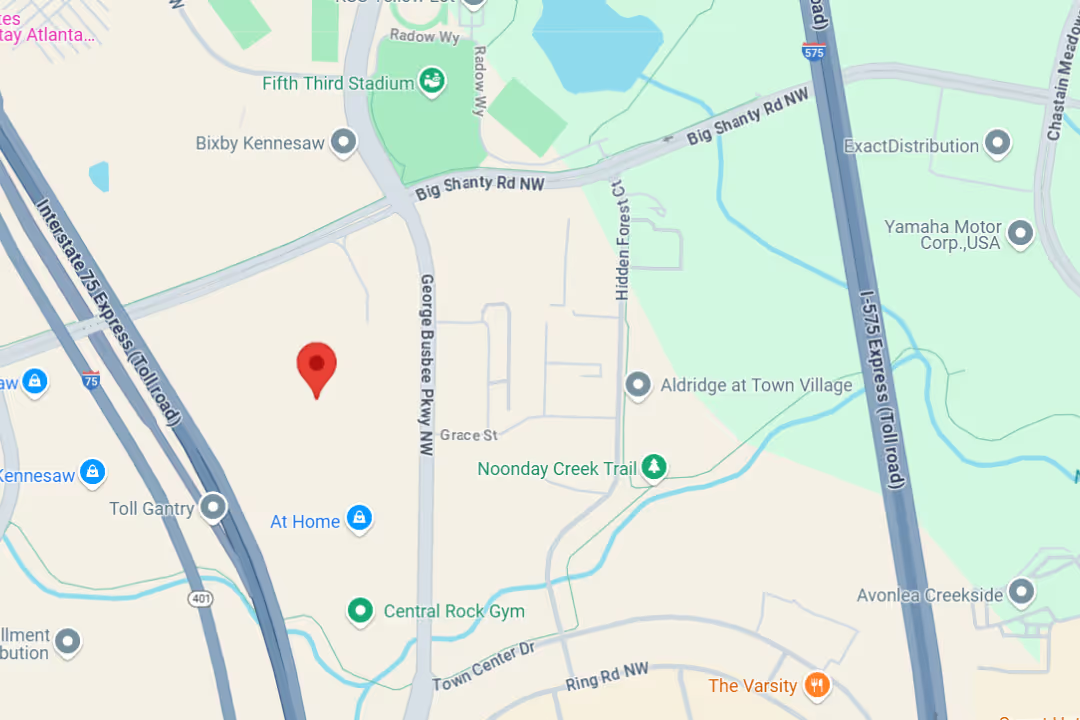
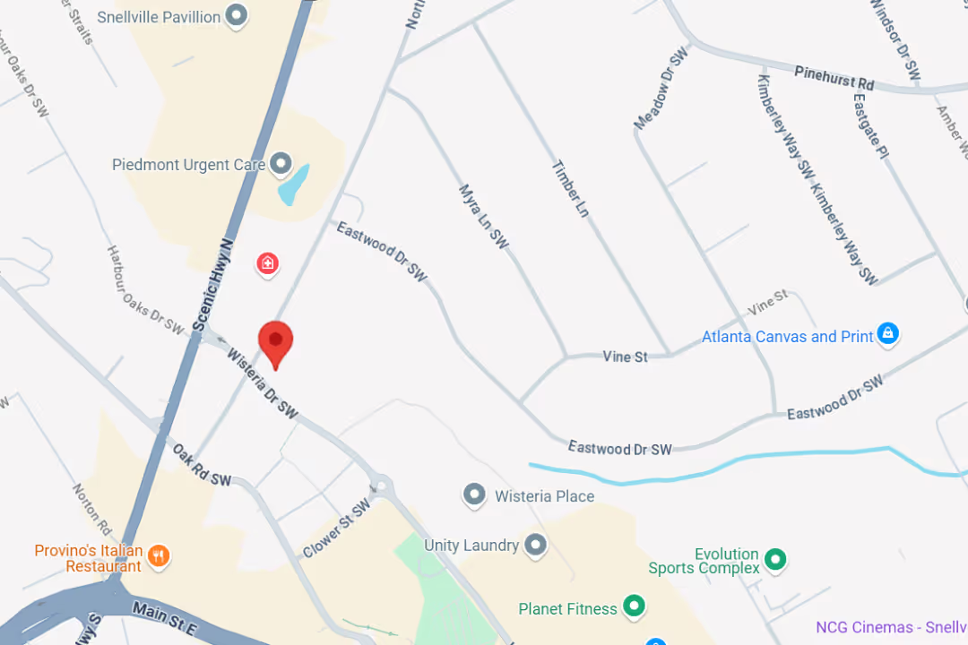
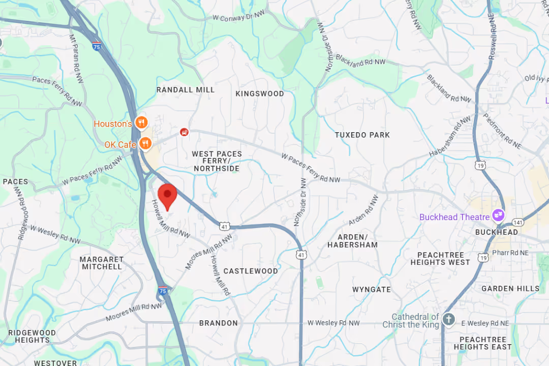
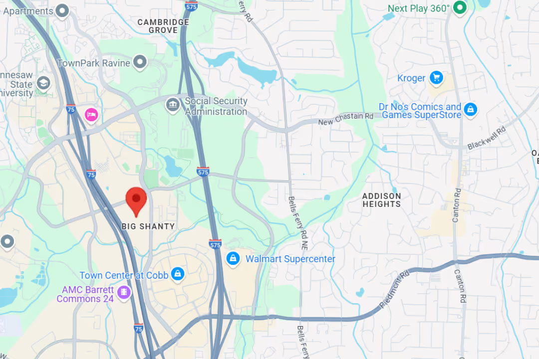
If you are struggling with lower back or radiating leg pain, do not wait for the symptoms to worsen. You can schedule a comprehensive evaluation at any of our convenient Metro Atlanta locations, including Snellville, Kennesaw, Marietta, and Buckhead. You can also explore our Lumbar Radiculopathy (Pinched Nerve) Condition Guide to learn more about how we can help you live pain-free.
At Medici, you’re more than your MRI.
We take time to hear your story, understand your pain, and create a plan that actually works for you.

Our team delivers specialist care at convenient locations across Metro Atlanta:
Get expert tips on injury recovery, pain relief, joint health, and movement strategies—straight from our Fellowship-Trained team.